Quality of life related to health and clinical results after radiation therapy of patients with intracranial meningioma
By Dominik Lisowski, Jannik Trömel, Paul Lutyj, Victor Lewitzki, Philipp E. Hartrampf, Bülent Polat, Michael Flentje & Jörg Tamihardja
Summary
This retrospective study, conducted in a single establishment, focused on long -term result, toxicity and quality of life related to health (QVLS) in patients with meningioma after radiotherapy.
We have analyzed the data of 119 patients who received radiotherapy in our service between 1997 and 2014 for an intracranial meningiom of I-III of the WHO. Fractional stereotaxic radiotherapy (FSRT), intensity modulation radiotherapy (IMRT) or radiosurgical radiation have been applied.
Les questionnaires EORTC QLQ-C30 et QLQ-BN20 ont été remplis pour l’évaluation de la QVLS. La survie globale (OS) pour l’ensemble du groupe d’étude était de 89,6 % à 5 ans et de 75,9 % à 10 ans. Le contrôle local (CL) à 5 et 10 ans était de 82,4% et 73,4%, respectivement. Une récidive locale a été observée chez 22 patients (18,5 %). Des toxicités aiguës et chroniques de plus haut grade ont été observées chez sept patients (5,9%) et cinq patients (4,2%), respectivement. L’état de santé global a été évalué avec une moyenne de 59,9 points (SD 22,3) sur le QLQ-C30. En conclusion, la radiothérapie a permis d’obtenir de très bons taux de survie à long terme et de contrôle de la tumeur avec de faibles taux de toxicités sévères mais avec une détérioration de la QVLS à long terme.
Les méningiomes sont des néoplasmes dérivés des cellules arachnoïdiennes des leptoméninges et sont les tumeurs intracrâniennes primaires les plus fréquentes chez les adultes avec 15 à 30 % (1). Les femmes sont deux fois plus souvent touchées que les hommes, cependant, les hommes ont tendance à développer des formes plus agressives de méningiomes (2). Les méningiomes sont classés en trois groupes par l’Organisation mondiale de la santé (OMS) en fonction de leurs caractéristiques histologiques.
Environ 80 à 85 % de tous les méningiomes sont classés dans la catégorie des méningiomes non malins (grade I de l’OMS), qui présentent généralement un taux de croissance lent et une expansion non invasive. Seulement 5 à 15 % des méningiomes sont considérés comme des méningiomes atypiques (grade II de l’OMS) et seulement 1 à 3 % sont des méningiomes malins (grade III de l’OMS) avec une tendance à l’invasion du cerveau (3,4).
Une nouvelle classification des méningiomes est basée sur des marqueurs moléculaires pour prédire les résultats cliniques avec plus de précision (5). Le traitement multimodal des méningiomes dépend du classement de l’OMS ainsi que de l’état de la résection et peut inclure la chirurgie, la radiothérapie, la thérapie par radionucléides à récepteurs peptidiques (PRRT) ou l’attente vigilante (6).
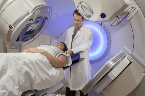
La radiothérapie est généralement appliquée comme traitement adjuvant ou en cas de rechute. En cas de méningiome non résécable, la radiothérapie primaire est l’option thérapeutique la plus courante. Elle peut être conduite en termes de radiothérapie à modulation d’intensité (IMRT), de radiothérapie stéréotaxique fractionnée (FSRT) ou de radiochirurgie (7). Bien que les techniques modernes d’irradiation aient diminué la quantité et la gravité de la toxicité aiguë et tardive, des publications antérieures ont observé des effets indésirables de haut niveau après une radiothérapie du cerveau, tels qu’un déficit du champ visuel, une neuropathie, une nécrose cérébrale, une dysfonction hypophysaire et des événements vasculaires cérébraux (8,9,10,11,12,13).
Les troubles cognitifs, les pertes de mémoire et les changements de personnalité peuvent être objectivement difficiles à quantifier, mais ont un impact énorme sur la vie quotidienne des individus (9,14). Par conséquent, tout effet secondaire aigu ou tardif peut entraîner une détérioration significative de la qualité de vie liée à la santé (QVLS) (15). Jusqu’à présent, peu de données ont été publiées sur la toxicité et la QVLS après radiothérapie chez les patients atteints de méningiome. Par conséquent, la présente analyse rétrospective monocentrique visait à fournir des données sur la QVLS, les effets secondaires et l’efficacité à long terme après une radiothérapie chez un grand groupe de patients atteints de méningiome.
Methods
Nous avons analysé rétrospectivement les données de 119 patients consécutifs atteints de méningiome qui ont été traités dans notre service entre 1997 et 2014. L’approbation éthique a été levée par le comité d’éthique local de l’Université de Wuerzburg en raison de la nature rétrospective de l’étude et toutes les procédures effectuées faisaient partie des soins de routine. Toutes les méthodes ont été réalisées conformément aux normes éthiques du comité de recherche de l’établissement et à la déclaration d’Helsinki de 1964 et ses amendements ultérieurs ou à des normes éthiques comparables.
The informed consent was obtained from all the subjects and/or their legal tutor (s) before planning of treatment. The main evaluation criterion of our study was the quality of life, evaluated by the main questionnaire on the quality of life of the European organization for the research and treatment of cancer (EORTC) (QLQ-C30) version 3.0 and the questionnaire of the module on the brain cancer of the EORTC (QLQ-BN20).
The secondary evaluation criteria were toxicity linked to treatment, local control (CL) at 5 and 10 years and overall survival (SG). Local control was defined as the time elapsed between the start of the irradiation and the appearance of the first progression on the site treated with imaging. Overall survival has been defined as time elapsed between diagnosis and the last documented follow -up or death, whatever the cause. We have been included all patients with meningioma who were treated with radiotherapy during the given period and who had no sign of spinal infiltration. In the event of multiple treatment series, we have analyzed a single series.
Patients with history of another cancer, regardless of the previous processing, were included in the database. All cases were discussed in an interdisciplinary neuro-oncological revision committee before treatment.
Treatment planning
For normofractional radiotherapy, the raw tumor volume (GTV) has been extended from 8 to 15 mm, depending on the WHO grade and the location of the tumor, to generate the clinical target volume (CTV). The CTV was extended by 3 mm to obtain the Target Planning Volume (PTV). The dose was prescribed at the average Dose of the PTV. In case of stereotaxic radiotherapy, a margin of 1 to 2 mm was added to the GTV for the PTV. The IMRT was administered according to the STEP-AND-SHOOT technique with 3 to 9 fields or according to the VMAT technique (Volumetric Intensity Modulated Arc Therapy) with two dynamic arcs. All radiotherapies were made with photon bundles using a linear Elekta Synergy® or Siemens Primus accelerator. The GTV has been delimited on computer assisted tomography (TAO) with magnetic resonance imaging (MRI) coinciding with Pinnacle (3) (Philips Radiation Oncology Systems, Fitchburg, Wi, USA).
Follow up
Clinical and radiological follow -up including an MRI with contrast was carried out 6 to 12 weeks after radiotherapy, then once or twice a year, unless an earlier examination is envisaged due to a suspicion of relapse. Imaging examinations were evaluated by two (neuro) independent radiologists. The dimensions of the tumor have been measured from an axial MRI sequence weighted in T1 with contrast or a scanner with contrast. In the event of a multifocal presence, the location of the tumor was defined by the site of the largest lesion. The RANO criteria (Responses Assessment in Neuro-ONCOLOGY) were used to assess the progression of the tumor (16). The location of the tumor was classified in the base of the skull, the cerebral falx, the hemispherical convexity or the sheath of the optic nerve.
The clinical examination included the evaluation of the neurological state. For the assessment of acute and late toxicity, version 5.0 of Common Terminology Criteria for opposing events (CTCAE) has been used. Acute toxicity was evaluated up to 90 days after the end of the irradiation. For the evaluation of the quality of life, the questionnaires EORTC QLQ-C30 version 3.0 and EORTC QLQ-BN20 were filled during follow-up visits or sent to patients. The Karnofsky Performance Status (KPS) was evaluated before the treatment and at the time of the QVLS assessment. The data from the QLQ-C30 and QLQ-BN20 have been compared to the already published data of historic cohorts (17,18,19,20,22,23).
Statistics
All data was analyzed with IBM SPSS Statistics 26.0. The statistical significance threshold has been set at a bilateral P <0.05. We assumed that the data of the study carried out were missing at random (Mar). As very few values were missing, we used the suppression in pairs to maintain a sufficient sample size and power. With regard to questionnaires QLQ-C30 and BN20, a relevant clinical difference was defined when the point difference was greater than 10 points (24.25). The SG and the LC were calculated using Kaplan-Meier statistics. The Log-Rank test was used to determine the statistical significance of the difference in SG or CL between the different groups. For multivariate analysis, a regression of proportional risk of COX has been carried out. Mann-Whitney-U and Kruskal-Wallis tests were carried out due to parameters not normally distributed according to the Shapiro-Wilk test. To correlate the grades of toxicity with the characteristics of the treatment, the location of the tumor and the QVLS data, the meaning of the Kendall Tau-B correlation coefficient has been evaluated. Multiple linear regressions have been used to find confusion factors for overall health.
Results
Treatment results
Meningioma was histologically determined in 76 patients (63.9%). In 43 patients (36.1%), the diagnosis was based on radiological signs after examination by magnetic resonance imaging (MRI) by at least two (neuro) radiologists. Irradiation was administered in 56 patients (47.0 %) during the initial diagnosis, 41 patients (34.5 %) having been irradiated in the year following the diagnosis. The other 63 patients (52.9%) were treated at the time of the relapse of meningioma. In total, 37 patients received primary radiotherapy due to the non-development of meningioma or inoperability that was conditioned by age and comorbidities. The median age of patients who received primary, adjuvant and recurrent radiotherapy was 70.2, 58.4 and 56.7 years, respectively. The median axial size of the tumor was 2.5 cm (IQR 1.5-3.8 cm) in the longest orientation at the start of radiotherapy. FSRT, IMRT and radiosurgery alone were carried out in 67 (56.3 %), 48 (40.3 %) and four (3.3 %) patients, respectively. Sequential extra irradiation was administered in 38 patients (31.9 %). A total median dose of 54.0 Gy (IQR 54.0-58.5 Gy), 60.0 Gy (IQR 54-61.2 Gy) and 60.0 Gy (IQR 59.4-60.3 Gy) was administered for the MEMB MES MS I, II and III, respectively. For stereotaxic radiotherapy, a total median dose of 19.5 Gy (interval 17.5-21 Gy) was prescribed at the 68 % PTV encompassing the isodosis. A patient was treated by an entire brain irradiation at 30.0 Gy and a sequential boost on the lesion of meningioma at 15.0 Gy. In 26 patients (21.8 %), Imaging by post-emission (TEP) to (68) Ga-Dota0-Phe1-Tyr3 Octreotide (Dotatoc), at (68) OCTREOTATE (Dotatate) or (18) F-Fluoroethyl-L-Tyrosine (FET) was carried out and merged to improve the definition of the target volume. Nine patients (7.6 %) received an additional RTPE using (177) Lu-Dotatoc with an average dose of 7.5 Gy (standard deviation ± 0.3). Three patients received concomitant or sequential chemotherapy. The characteristics of all patients are summarized in Table 1.
Table 1 Characteristics of patients (n = 119).
Quality of life
As 37 patients (31.1 %) had already died at the time of the survey, 82 questionnaires were sent or sent, 49 of which were duly completed and returned, which gives a response rate of 59.8 %. A questionnaire was returned with the notification that the corresponding patient had died. The median KPS of the patients interviewed before radiotherapy and at the time of the QVLS evaluation was 90 respectively (interval 50-100) and 90 (interval 40-100). The median kps of the entire study group was 80 (fork 30-100). The questionnaires were filled in the median 4.8 years (IQR 2.7-9.2 years) after radiation therapy by patients of 64.4 years (IQR 59.0-72.5 years). Of the 49 responders, three received additional cranial radiotherapy and four sequential extracranial radiotherapy. Analysis of the basic characteristics of patients between the group of responders and that of non-repairrs was not significant, except for the PRRT (additional table S1).
In terms of self-assessment, the overall state of health was evaluated with an average of 59.9 points (SD 22.3) on the EORTC QLQ-C30 with functional scales ranging from an average of 55.6 to 71.2 points (Table 2). We were able to detect a relevant decrease on the functional scale for physical functioning, the role, cognitive and social functioning, which was accompanied by an increase on the scale of symptoms for fatigue, pain, dyspnea, insomnia, constipation and financial impact. There was no statistically significant correlation between the maximum grade of chronic toxicity and fatigue (p = 0.41), nausea and vomiting (p = 0.43), pain (p = 0.12), dyspnea (p = 0.5), insomnia (p = 0.35), loss of appetite (p = 0.20) 0.09), diarrhea (p = 0.49) and financial difficulties (p = 0.46). Sequential radiotherapy, secondary metachron, meningiomal secondary tumors were not confusion factors for overall health on QLQ-C30 (p ≥ 0.05). On the QLQ-BN20 EORTC, the most frequent disabilities were drowsiness, uncertainty about the future and weakness of the legs (Table 2). Compared to previous cohorts, our data showed partially less good results on the QLQ-C30 (Figure 1) and on the QLQ-BN20 (additional figure S1).
Table 2 Scores of the ITEMS du EORTC QLQ-C30 and BN20 (n = 49).
Table 1
(a) Comparison of the functional scales of the QLQ-C30 with the data published above. Higher scores in functional areas suggest a higher level of operation and better quality of life. Ql2 = global state of health (revised); PF2 = physical functioning (revised); RF2 = functioning of roles (revised); EF = emotional functioning; See = Cognitive operation; SF = social functioning. (b) Comparison of QLQ-C30 symptom scales with previously published data. Higher scores in the symptomatic fields suggest a lower level of operation and a worse quality of life. Abbreviations: FA = Fatigue; Nv = nausea and vomiting; Pa = pain; Dy = dyspnea; SL = insomnia; AP = loss of appetite; Co = constipation; Di = diarrhea; Fi = financial difficulties. *Unpublished data.
Side effects
Des toxicités aiguës liées aux radiations et ayant une signification clinique (grade CTCAE ≥ 3) ont été enregistrées chez sept patients (5,9 %). Il s’agissait notamment d’un cas d’amaurose avec altération préalable de la visibilité et de trois cas de vertiges.
The other toxicities of Grade CTCAE 3 were as follows: nausea, headache, irradiation dermatitis, fatigue and mucosite. In two cases, the irradiation had to be interrupted due to a deterioration of the general state. Acute side effects of grade 1 and 2 occurred in 52.9% and 37.8% of cases, respectively. Fatigue, alopecia, headache, radical dermatitis, dizziness, as well as nausea and vomiting were the most frequently reported acute side effects. In 3.4 % of cases, no adverse effects have been reported.
Severe chronic toxicities (CTCAE grade ≥ 3) were observed in five patients (4.2 %). There was a case of overditas (grade CTCAE 4) and a case of amaurosis (grade CTCAE 4) with an insufficiency of the anterior pituitary gland (grade CTCAE 3). The other three cases suffered from fatigue, exhaustion, confusion or headache (all of grade ctcae 3). Chronic CTCAE 1 and 2 chronic side effects were observed in 11.8 % and 20.2 % of patients, respectively. The most frequent chronic side effect was chronic headache, which occurred in 7.5 % of cases. In addition, a circumscribed toxicity of the SNC (6.7 %), memory and concentration disorders (5.9 %) as well as fatigue (5.8 %) were relatively frequent.
All average dose values for risk organs in our study group were lower than the recommended limits. Patients with grade toxicities ≤ 2 received an average total dose of 54.9 Gy. The average total dose in patients with grade toxicities ≥ 3 was 57.9 gy. There was no statistically significant correlation between the maximum toxicity grade and the total dose (p = 0.55), PTV (p = 0.86) GTV (p = 0.52) or the location of the tumor (p = 0.56). There was a statistically significant correlation between the grade of toxicity of acute fatigue and the scope of QLQ-C30 fatigue symptoms (p = 0.03).
Local control
Median follow-up was 5.4 years (IQR 2.9-9.7 years). The estimated rates of CL at 5 and 10 years were 82.4 % and 73.4 %, respectively (fig. 2A). The median time until recurrence was not reached at the time of data analysis. In total, 22 patients (18.5 %) had a relapse in the field, three patients with MEMB MESGI MENINGIOME, SIX MENINGIOMA OF GRADE II of WHO and eight MENINGIOMA OF GRADE III of WHO. A relapse also occurred in five patients without meningioma confirmed histologically. A patient with highly suspected type II neurofibromatosis was diagnosed with a relapse of meningioma twice. The histological grade was significant and suggestive to influence the LC in the univariate (p <0.001) and multivariate (p = 0.05) analysis, respectively (fig. 2b). The rank of Simpson (i-iii vs. iv-v) had no statistically significant impact on the cl. The location of the tumor (p = 0.032) as well as the GTV for the patient's subgroup with OMS II and III grade meningiomas (p = 0.023) were significant in univariate analysis, but not in multivariate analysis. No significant difference of CL could be observed by comparing a cumulative dose of ≥ 60 Gy versus <60 Gy for all patients (p = 0.37) or for patients with MEMBS II and III grade meningiomas (p = 0.46).
Table 2
Local control shown by Kaplan-Meier analysis for all patients (A) and laminate according to the WHO ranking (B). The WHO classification was suggestive to influence local control (p = 0.05). Global survival is represented by an analysis of Kaplan-Meier for all patients (C) and laminate according to the WHO classification (D). The WHO classification was highly significant for global survival (p = 0.002).
Global survival
Au total, 38 patients sont décédés au moment de l’enquête. Sur les 38 décès, 16 patients (42,1%) ont été présumés décédés de la maladie du méningiome et dix patients (26,3%) ont succombé à des comorbidités. Dans 12 cas (31,6 %), la cause du décès n’était pas claire. La SG estimée à 5 et 10 ans était de 89,6 % et 75,9 %, respectivement (Fig. 2c). La SG médiane était de 17,5 ans. Les taux de survie différaient significativement selon le grade OMS (p = 0,002). Le KPS (≥ 90 % vs. < 90 %) (p = 0,046), le GTV (p = 0,001), le moment de la radiation (p = 0,005) et l’âge (p = 0,001) ont eu un impact significatif sur la SG en analyse univariée. Après une analyse multivariée, le grade OMS (p = 0,002) et le GTV (p = 0,001) restaient significatifs pour la SG (Fig. 2d). La SG n’était pas significativement affectée par le sexe, le grade de Simpson, la localisation de la tumeur et le volume tumoral avant traitement. Comme pour la CL, aucune différence significative de la SG n’a été trouvée en comparant les radiothérapies avec une escalade de dose supérieure à 60 Gy pour tous les patients (p = 0,32) ou pour les patients avec des méningiomes de grade OMS II et III (p = 0,08).
Discussion
A notre connaissance, cette étude est la première à évaluer les données de HRQoL en utilisant les questionnaires QLQ-C30 et BN20 pour des patients exclusivement atteints de méningiome et ayant reçu une radiothérapie. Notre recherche dans la base de données n’a permis de trouver que quelques publications évaluant la QVLS à l’aide des questionnaires QLQ-C30 et BN20 chez des patients atteints de méningiome, la plupart sous forme d’analyses de sous-groupes non planifiées (17,18,19,23). Le taux de réponse pour l’évaluation de la QDV était de 59,8 %. Nous n’avons pas pu détecter une raison spécifique pour le retour manquant des 32 questionnaires et nous ne pouvons que spéculer sur la raison pour laquelle le taux de réponse était limité. Bien que nous n’ayons pas pu trouver de différence significative entre les groupes ayant répondu et n’ayant pas répondu, sauf pour la PRRT (tableau supplémentaire S1), un biais de sélection ne peut être exclu en raison du taux de réponse limité. Nos données montrent des résultats légèrement inférieurs en matière de QVLS par rapport aux données déjà publiées pour les patients atteints de méningiome, bien que la comparabilité puisse être limitée en raison des différentes méthodes d’acquisition des données et de la composition des groupes de patients (26). Par exemple, Erharter et al. ont effectué une présélection des patients en excluant les patients présentant des troubles cognitifs sévères, ce qui a donné lieu à des scores de QVLS plus élevés (23). Shin et al. n’ont fourni aucune information supplémentaire sur la composition des groupes de patients atteints de méningiome (19). Budrukkar et al. ont évalué la QVLS dans un sous-groupe de patients atteints de tumeurs cérébrales bénignes, qui n’était pas limité aux seuls patients atteints de méningiomes (18). Konglund et al. ont rapporté des scores QLQ-C30 plus élevés, ce qui est probablement attribuable à des différences de groupe car leur cohorte était composée à 94% de méningiomes bénins réséqués sans radiothérapie (17). La radiothérapie primaire est souvent choisie pour les tumeurs avancées et inopérables et les méningiomes bénins ne devraient pas être irradiés après une résection complète selon les directives de l’Association européenne de neuro-oncologie (EANO) (27,28).
The heterogeneity of our examination group examined must be taken into account. Our study group was mainly composed of patients with unfavorable tumor location and 100 patients received radiotherapy as first -line treatment or at the time of relapse. Recurrent or incompletely resonated meningiomas are likely to have less good results, with more clinically significant side effects and, therefore, a lower QVLS (13). The grade II (16.8 %) and III (15.1 %) meningioma rate of the WHO in our study was higher than the average, which leads to the over -representation of high grade meningiomas (Grade II and III of WHO). In addition, 21 patients (17.6 %) of our study group reported another malignant tumor before the QDV assessment, which could be a factor of confusion.
La QDV a été déterminée dans un délai médian de 4,8 ans après le traitement dans notre étude, ce qui laisse la possibilité que d’autres maladies influençant négativement la QDV soient des facteurs de confusion, comme un accident vasculaire cérébral ou une détérioration cognitive due au vieillissement. L’absence d’évaluation longitudinale de la QVLS est un facteur limitant de notre analyse, car les données sur la QVLS n’ont été évaluées qu’à un moment précis du suivi. Il manque donc une enquête préalable au traitement pour comparer la QVLS et identifier les facteurs de confusion possibles ou les sous-groupes de patients présentant une plus forte détérioration de la QVLS. Étant donné que seule la QVLS à long terme a été évaluée dans notre étude, les effets bénéfiques directement après la radiothérapie ou la chirurgie entraînant des gains fonctionnels et une meilleure QVLS n’ont pas été mesurés, contrairement aux études de Budrukkar et al, Konglund et al et Bitterlich et al. (17,18,29).
Les toxicités aiguës sévères évaluées par les médecins ne sont apparues que dans 5,9% des cas, confirmant que la radiothérapie a des effets secondaires légers lorsqu’elle est appliquée chez les patients atteints de méningiome. Le seul cas d’amaurose aiguë pourrait être lié à la croissance de la tumeur, car le patient souffrait d’une grave déficience visuelle avant la radiothérapie et a reçu une radiation palliative à une dose plus faible. Bien que 36,2 % des patients aient signalé des toxicités chroniques, seuls 4,2 % ont souffert d’un effet secondaire chronique de grade CTCAE ≥ 3. Nos résultats sont conformes aux données publiées précédemment en termes de toxicités aiguës et tardives (0-49,9 %) (7,10,12,14,30,31).
Our median dose of applied radiation was comparable to that of existing literature. Based on the data already published, a dose of 54-60 GY is indicated and well tolerated for the MEMB MES MOS. In our Cohort of Méningiomes de Grade I de l'OMS, a dose up to 66.0 Gy was accepted if histopathological specimens had angiomatous or fibrous components. For high grade meningiomas, a total median dose of 60.0 Gy was prescribed in our study. A minimum dose of 60 Gy is generally prescribed for MENINGIOMS OF GRADE III of WHO to ensure long -term local control (32.33). The prescription of the dose for the MENINGIOMS of Grade II of the WHO, however, is not consistent in the literature. Depending on the state of resection, high -dose irradiation of 60 Gy or 70 Gy has been prescribed for all patients with Grade II meningiomas in the EORTC 22042 study, while patients with grade II meningiomas of WHO newly diagnosed with total macroscopic resection of 54 gy in the RTOG 053934.35 study. Survival without progression (SSP) and the SG at three years were comparable in both studies. The long -term results of the two studies have not yet been published. In retrospective analyzes, the increase in dose is however associated with an improvement in the clinical result and can be prescribed for the MEM Grade II meningiomas (32,33,36,37).
The existing reports on the factors influencing the SG and the CL for meningiomas are inconsistent, except for the rank of the WHO (11,15,30,38,39,40,41). In accordance with these results, our data confirmed that the rank of WHO had a significant impact on the SG in univariate and multivariate analysis and also affected local control. Due to the lack of studies with a large number of patients, statistics for the SG and CL in the MENINGIOMS of Grade II and III of the WHO show a wide variance (0.0-89.0%) (additional table S2) (30,32,33,38,40,42,43). Our CL estimated at 5 years for MENINGIOMS of Grade II of WHO (66.7%) and Grade III of WHO (53.1%) is compatible with the majority of published data (additional Table S2) (11,13,15,30,32,33,38,40,42,45,47). Our survival rates at 5 years and 10 years for each OMS grade, however, seem more favorable than those published. This could be due to the fact that our low number of patients with high -risk meningiomas limits statistical information. In addition, the histological classification of older samples has not been updated according to the revised classification system of WHO since 2016, which has influenced the indication of radiotherapy, the target volume, the applied dose and probably the result (48). Although the concordance for the histopathological classification of meningiomas is relatively high, there are still divergences between observers and between institutions, which could cause a bias in the results (49).
Conclusion
In our cohort of patients with meningiomas, mostly at an advanced or relapse stage, radiotherapy has shown an excellent prognosis with regard to SG and LC and an acceptable QVLS with low toxicity reported by doctors. The deterioration of the QDV must be considered in relation to the risk of recurrence of meningioma and can therefore guide decision -making in favor of or not radiotherapy. Prospective studies should aim to improve QVLS without deteriorating the oncological result.
Data availability
The data sets generated and/or analyzed during this study are available from the corresponding author on reasonable request.
References
- Bondy, M. & Ligon, B. L. Epidemiology and etiology of intracranial meningiomas: A review. J. Neurooncol. 29, 197–205. https://doi.org/10.1007/BF00165649 (1996).
Article CAS PubMed Google Scholar
- Perry, A., Stafford, S. L., Scheithauer, B. W., Suman, V. J. & Lohse, C. M. Meningioma grading: an analysis of histologic parameters. Am. J. Surg. Pathol. 21, 1455–1465. https://doi.org/10.1097/00000478-199712000-00008 (1997).
Article CAS PubMed Google Scholar
- Kleihues, P. & Cavenee, W. K. Pathology and Genetics of Tumours of the Nervous System (International Agency for Research on Cancer, 2000).
- Dolecek, T. A. et al. Epidemiology of meningiomas post-public law 107–206: The benign brain tumor cancer registries amendment act. Cancer 121, 2400–2410. https://doi.org/10.1002/cncr.29379 (2015).
- Nassiri, F. et al. A clinically applicable integrative molecular classification of meningiomas. Nature 597, 119–125. https://doi.org/10.1038/s41586-021-03850-3 (2021).
Article ADS CAS PubMed Google Scholar
- Hartrampf, P. E. et al. Long-term results of multimodal peptide receptor radionuclide therapy and fractionated external beam radiotherapy for treatment of advanced symptomatic meningioma. Clin. Transl. Radiat. Oncol. 22, 29–32. https://doi.org/10.1016/j.ctro.2020.03.002 (2020).
Article PubMed PubMed Central Google Scholar
- Gondi, V., Tome, W. A. & Mehta, M. P. Fractionated radiotherapy for intracranial meningiomas. J. Neurooncol. 99, 349–356. https://doi.org/10.1007/s11060-010-0368-5 (2010).
- Al-Mefty, O., Kersh, J. E., Routh, A. & Smith, R. R. The long-term side effects of radiation therapy for benign brain tumors in adults. J.Neurosurg. 73(4), 502–512. https://doi.org/10.3171/jns.1990.73.4.0502 (1990).
Article CAS PubMed Google Scholar
- Miralbell, R. et al. The role of radiotherapy in the treatment of subtotally resected benign meningiomas. J. Neuro-Oncol. 13, 157–164. https://doi.org/10.1007/BF00172765 (1992).
- Goldsmith, B. J., Wara, W. M., Wilson, C. B. & Larson, D. A. Postoperative irradiation for subtotally resected meningiomas: A retrospective analysis of 140 patients treated from 1967 to 1990. J. Neurosurg. 80(2), 195–201. https://doi.org/10.3171/jns.1994.80.2.0195 (1994).
Article CAS PubMed Google Scholar
- Debus, J. et al. High efficacy of fractionated stereotactic radiotherapy of large base-of-skull meningiomas: long-term results. J. Clin. Oncol. Offl. J. Am. Soc. Clin. Oncol. 19, 3547–3553. https://doi.org/10.1200/JCO.2001.19.15.3547 (2001).
- Barbaro, N. M. et al. Radiation therapy in the treatment of partially resected meningiomas. Neurosurgery 20, 525–528. https://doi.org/10.1227/00006123-198704000-00003 (1987).
Article CAS PubMed Google Scholar
- Taylor, B. W. Jr., Marcus, R. B. Jr., Friedman, W. A., Ballinger, W. E. Jr. & Million, R. R. The meningioma controversy: Postoperative radiation therapy. Int. J. Radiat. Oncol. Biol. Phys. 15, 299–304. https://doi.org/10.1016/s0360-3016(98)90008-6 (1988).
- Nutting, C. et al. Radiotherapy in the treatment of benign meningioma of the skull base. J. Neurosurg. 90, 823–827. https://doi.org/10.3171/jns.1999.90.5.0823 (1999).
Article CAS PubMed Google Scholar
- Combs, S. E. et al. Skull base meningiomas: Long-term results and patient self-reported outcome in 507 patients treated with fractionated stereotactic radiotherapy (FSRT) or intensity modulated radiotherapy (IMRT). Radiother. Oncol. J. Eur. Soc. Ther. Radiol. Oncol. 106, 186–191. https://doi.org/10.1016/j.radonc.2012.07.008 (2013).
- Huang, R. Y. et al. Proposed response assessment and endpoints for meningioma clinical trials: Report from the Response Assessment in Neuro-Oncology Working Group. Neuro Oncol. 21, 26–36. https://doi.org/10.1093/neuonc/noy137 (2018).
Article PubMed Central Google Scholar
- Konglund, A. et al. Outcome following surgery for intracranial meningiomas in the aging. Acta Neurol. Scand. 127, 161–169. https://doi.org/10.1111/j.1600-0404.2012.01692.x (2013).
Article CAS PubMed Google Scholar
- Budrukkar, A. et al. Prospective assessment of quality of life in adult patients with primary brain tumors in routine neurooncology practice. J. Neurooncol. 95, 413–419. https://doi.org/10.1007/s11060-009-9939-8 (2009).
- Shin, Y. S. & Kim, J. H. Validation of the Korean version of the European Organization for Research and Treatment of Cancer brain cancer module (EORTC QLQ-BN20) in patients with brain tumors. Health Qual. Life Outcomes 11, 145. https://doi.org/10.1186/1477-7525-11-145 (2013).
Article PubMed PubMed Central Google Scholar
- Schwarz, R. & Hinz, A. Reference data for the quality of life questionnaire EORTC QLQ-C30 in the general German population. Eur. J. Cancer 37, 1345–1351. https://doi.org/10.1016/s0959-8049(00)00447-0 (2001).
Article CAS PubMed Google Scholar
- Nolte, S. et al. General population normative data for the EORTC QLQ-C30 health-related quality of life questionnaire based on 15,386 persons across 13 European countries, Canada and the Unites States. Eur. J. Cancer 107, 153–163. https://doi.org/10.1016/j.ejca.2018.11.024 (2019).
Article CAS PubMed Google Scholar
- Neil W Scott, PMF, Neil K Aaronson, Andrew Bottomley, Alexander de Graeff, Mogens Groenvold, Chad Gundy, Michael Koller, Morten A Petersen, Mirjam Ag Sprars. (Ed Eortc Groups) (2008).
- Erharter, A. et al. Implementation of computer-based quality-of-life monitoring in brain tumor outpatients in routine clinical practice. J. Pain Symptom Manage. 39, 219–229. https://doi.org/10.1016/j.jpainsymman.2009.06.015 (2010).
- King, M. T. The interpretation of scores from the EORTC quality of life questionnaire QLQ-C30. Qual. Life Res. Int. J. Qual. Life Aspects Treatment Care Rehabil. 5, 555–567. https://doi.org/10.1007/BF00439229 (1996).
- Osoba, D., Rodrigues, G., Myles, J., Zee, B. & Pater, J. Interpreting the significance of changes in health-related quality-of-life scores. J. Clin. Oncol. Offl. J. Am. Soc. Clin. Oncol. 16, 139–144. https://doi.org/10.1200/JCO.1998.16.1.139 (1998).
- Meskal, I., Gehring, K., Rutten, G. J. & Sitskoorn, M. M. Cognitive functioning in meningioma patients: A systematic review. J. Neurooncol. 128, 195–205. https://doi.org/10.1007/s11060-016-2115-z (2016).
Article PubMed PubMed Central Google Scholar
- Goldbrunner, R. et al. EANO guidelines for the diagnosis and treatment of meningiomas. Lancet Oncol. 17, e383-391. https://doi.org/10.1016/S1470-2045(16)30321-7 (2016).
- Zamanipoor Najafabadi, A. H. et al. Impaired health-related quality of life in meningioma patients—A systematic review. Neuro-Oncol. 19, 897–907. https://doi.org/10.1093/neuonc/now250 (2017).
- Bitterlich, C. & Vordermark, D. Analysis of health-related quality of life in patients with brain tumors prior and subsequent to radiotherapy. Oncol. Lett. 14, 1841–1846. https://doi.org/10.3892/ol.2017.6310 (2017).
Article PubMed PubMed Central Google Scholar
- Milker-Zabel, S. et al. Fractionated stereotactic radiotherapy in patients with benign or atypical intracranial meningioma: Long-term experience and prognostic factors. Int. J. Radiat. Oncol. Biol. Phys. 61, 809–816. https://doi.org/10.1016/j.ijrobp.2004.07.669 (2005).
- Minniti, G., Amichetti, M. & Enrici, R. M. Radiotherapy and radiosurgery for benign skull base meningiomas. Radiat. Oncol. 4, 42. https://doi.org/10.1186/1748-717X-4-42 (2009).
Article PubMed PubMed Central Google Scholar
- Boskos, C. et al. Combined proton and photon conformal radiotherapy for intracranial atypical and malignant meningioma. Int. J. Radiat. Oncol. Biol. Phys. 75, 399–406. https://doi.org/10.1016/j.ijrobp.2008.10.053 (2009).
- Hug, E. B. et al. Management of atypical and malignant meningiomas: Role of high-dose, 3D-conformal radiation therapy. J. Neurooncol. 48, 151–160. https://doi.org/10.1023/a:1006434124794 (2000).
Article CAS PubMed Google Scholar
- Weber, D. C. et al. Adjuvant postoperative high-dose radiotherapy for atypical and malignant meningioma: A phase-II parallel non-randomized and observation study (EORTC 22042–26042). Radiother. Oncol. J. Eur. Soc. Therap Radiol. Oncol. 128, 260–265. https://doi.org/10.1016/j.radonc.2018.06.018 (2018).
- Rogers, L. et al. Intermediate-risk meningioma: initial outcomes from NRG Oncology RTOG 0539. J. Neurosurg. 129, 35–47. https://doi.org/10.3171/2016.11.JNS161170 (2018).
- Kaur, G. et al. Adjuvant radiotherapy for atypical and malignant meningiomas: A systematic review. Neuro Oncol. 16, 628–636. https://doi.org/10.1093/neuonc/nou025 (2014).
Article PubMed PubMed Central Google Scholar
- McDonald, M. W. et al. Proton therapy for atypical meningiomas. J. Neurooncol. 123, 123–128. https://doi.org/10.1007/s11060-015-1770-9 (2015).
Article CAS PubMed Google Scholar
- Pasquier, D. et al. Atypical and malignant meningioma: outcome and prognostic factors in 119 irradiated patients. A multicenter, retrospective study of the Rare Cancer Network. Int. J. Radiat. Oncol. Biol. Phys. 71, 1388–1393. https://doi.org/10.1016/j.ijrobp.2007.12.020 (2008).
- Durand, A. et al. WHO grade II and III meningiomas: A study of prognostic factors. J. Neurooncol. 95, 367–375. https://doi.org/10.1007/s11060-009-9934-0 (2009).
- Adeberg, S. et al. Long-term outcome after radiotherapy in patients with atypical and malignant meningiomas–clinical results in 85 patients treated in a single institution leading to optimized guidelines for early radiation therapy. Int. J. Radiat. Oncol. Biol. Phys. 83, 859–864. https://doi.org/10.1016/j.ijrobp.2011.08.010 (2012).
- Piscevic, I. et al. The influence of adjuvant radiotherapy in atypical and anaplastic meningiomas: A series of 88 patients in a single institution. World Neurosurg. 83, 987–995. https://doi.org/10.1016/j.wneu.2015.02.021 (2015).
- Stafford, S. L. et al. Meningioma radiosurgery: Tumor control, outcomes, and complications among 190 consecutive patients. Neurosurgery 49, 1029–1037. https://doi.org/10.1097/00006123-200111000-00001 (2001).
Article MathSciNet CAS PubMed Google Scholar
- Rogers, C. L. et al. High-risk meningioma: Initial outcomes from NRG oncology/RTOG 0539. Int. J. Radiat. Oncol. Biol. Phys. 106, 790–799. https://doi.org/10.1016/j.ijrobp.2019.11.028 (2020).
- Solda, F. et al. Long-term efficacy of fractionated radiotherapy for benign meningiomas. Radiother. Oncol. J. Eur. Soc. Therap. Radiol. Oncol. 109, 330–334. https://doi.org/10.1016/j.radonc.2013.10.006 (2013).
- Hamm, K. et al. Radiosurgery/stereotactic radiotherapy in the therapeutical concept for skull base meningiomas. Zentralbl. Neurochir. 69, 14–21. https://doi.org/10.1055/s-2007-992138 (2008).
Article CAS PubMed Google Scholar
- Tanzler, E., Morris, C. G., Kirwan, J. M., Amdur, R. J. & Mendenhall, W. M. Outcomes of WHO Grade I meningiomas receiving definitive or postoperative radiotherapy. Int. J. Radiat. Oncol. Biol. Phys. 79, 508–513. https://doi.org/10.1016/j.ijrobp.2009.11.032 (2011).
- Wegner, R. E. et al. Linear accelerator-based stereotactic radiotherapy for low-grade meningiomas: Improved local control with hypofractionation. J. Centr. Nerv. Syst. Dis. 11, 1179573519843880. https://doi.org/10.1177/1179573519843880 (2019).
- Combs, S. E., Schulz-Ertner, D., Debus, J., von Deimling, A. & Hartmann, C. Improved correlation of the neuropathologic classification according to adapted world health organization classification and outcome after radiotherapy in patients with atypical and anaplastic meningiomas. Int. J. Radiat. Oncol. Biol. Phys. 81, 1415–1421. https://doi.org/10.1016/j.ijrobp.2010.07.039 (2011).
- Rogers, C. L. et al. Pathology concordance levels for meningioma classification and grading in NRG Oncology RTOG Trial 0539. Neuro Oncol. 18, 565–574. https://doi.org/10.1093/neuonc/nov247 (2016).
Download references
Funding
This publication was supported by the free access publication fund from the University of Wuerzburg. The financing of the open access was made possible and organized by Projokt Deal.
Author information
Authors and affiliations
- Department of Radio-Oncology, Wuerzburg University Hospital, Josef-Schneider-Str. 11, 97080, Wuerzburg, Germany
Dominik Lisowski, Paul Lutyj, Victor Lewitzki, Bülent Polat, Michael Flenger & Jörg Tamihardja
- Department of Internal Medicine, Leopoldina Hospital in Schweinfurt, Schweinfurt, Germany
Jannik Trömel
- Department of Nuclear Medicine, Wuerzburg University Hospital, Wuerzburg, Germany.
Philipp E. Hartrampf
Contributions
Study design: MF, BP, J.TA. Preparation of equipment, data collection: DL, J.TR., PL, J.TA., PH, VL Analysis and interpretation of data: DL, J.TR. Writing of the manuscript: DL. All the authors contributed to the revision of the project and approved the submissive version.
Corresponding author
Correspondence with Dominik Lisowski.
Ethics
Competing interests
The authors do not declare any competing interest.
Additional information
Publisher's note
Springer Nature remains neutral with regard to jurisdictional claims in published maps and institutional affiliations.