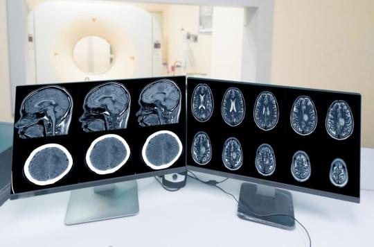
Opposed evolution of bone and soft parts of osteomeningioma associated with progesterone after stopping progesterone taking
https://pubmed.ncbi.nlm.nih.gov/36883659/
(translation made by the association, for good understanding by all)
Simona M. Florea, MD,1 Thibault Passeri, MD,1 Rosaria Abbritti, MD,1 Anne L. Bernat, MD,1, Sylvie Fontanel, MD,2 Isabelle Yoldjian, MD,3 Thomas Funck-Brentano, MD, PhD,4 Alain Weill, MD,5, Emmanuel Mandonnet, MD, PhD,1 and Sébastien Froelich, MD1
Départements de 1 neurochirurgie et 4 rhumatologie, Hôpital Lariboisière, AP-HP, Université de Paris ; 2 Agence régionale de santé Grand Est, Nancy ; 3 Agence nationale de sécurité du médicament et des produits de santé (ANSM), Saint-Denis ; et 5 Groupement d’intérêt scientifique EPI-PHARE, Saint-Denis, France.
ACP: Cyproterone acetate
OBJECTIF De nombreuses études ont confirmé l’existence d’un lien étroit entre les progestatifs et les méningiomes, ainsi que la régression et/ou la stabilisation des méningiomes après l’arrêt du traitement. Les ostéoméningiomes représentent un petit sous-groupe de méningiomes qui semblent être plus fréquents parmi les méningiomes liés aux progestatifs. Cependant, la spécificité du comportement de ce sous-groupe de méningiomes après l’arrêt du progestatif n’a pas encore été évaluée.
MÉTHODES Trente-six patients (âge moyen 49,5 ans) présentant au moins un ostéoméningiome lié à un progestatif (48 tumeurs au total) ont été identifiés à partir d’une base de données prospective de patients qui avaient été adressés à notre service pour un méningiome et dont l’utilisation d’acétate de cyprotérone, d’acétate de nomégestrol et/ou d’acétate de chlormadinone avait été documentée. Le traitement hormonal a été arrêté au moment du diagnostic pour tous les patients, et l’évolution clinique et radiologique de ce sous-groupe de tumeurs a été évaluée.
RÉSULTATS Pour la moitié des 36 patients, le traitement a été prescrit pour des signes d’hyperandrogénie, tels que l’hirsutisme, l’alopécie ou l’acné. La plupart des lésions étaient sphéno-orbitaires (35,4 %) ou frontales (31,2 %). Bien que la partie tissulaire du méningiome ait diminué dans 77,1 % des cas, la partie osseuse a présenté un comportement discordant avec 81,3 % de progression du volume. La combinaison d’œstrogènes, ainsi que la durée prolongée du traitement progestatif, semblent augmenter le risque de progression de la partie osseuse après l’arrêt du traitement (p = 0,02 et p = 0,028, respectivement). Aucune patiente n’a nécessité de traitement chirurgical au moment du diagnostic ou au cours de l’étude.
CONCLUSIONS Ces résultats montrent que si la partie intracrânienne molle de l’ostéoméningiome lié à la progestérone est la plus susceptible de régresser après l’arrêt du traitement, la partie osseuse est plus susceptible d’augmenter de volume. Ces résultats suggèrent la nécessité d’un suivi attentif de ces patients, en particulier ceux dont les tumeurs sont situées à proximité de l’appareil optique.
MOTS-CLÉS ostéoméningiome ; progestatif ; acétate de cyprotérone ; acétate de nomégestrol ; acétate de chlormadinone ; méningiome intra-osseux primaire ; oncologie ; tumeur
Les méningiomes sont des tumeurs bénignes du SNC qui représentent environ 39 % des tumeurs cérébrales primaires dans la population adulte.1 Au fil du temps, la relation entre les méningiomes et les hormones sexuelles a été bien établie et les arguments en faveur d’une telle association sont nombreux : prédominance féminine avec un ratio femme/homme de 2,3:1,1 accélération de la croissance pendant la grossesse et tendance au rétrécissement après l’accouchement, association signalée entre le carcinome mammaire et le méningiome,2-5 origine des méningiomes à partir des cellules arachnoïdiennes, qui expriment naturellement les récepteurs de la progestérone, ainsi que niveaux élevés d’expression des récepteurs de la progestérone et des androgènes dans les méningiomes.6,7 Des agents antiprogestérone tels que la mifépristone ont également été utilisés sur des méningiomes non résécables, avec un effet plutôt limité.8,9 Cependant, les mécanismes sous-jacents expliquant les relations entre les hormones sexuelles et les méningiomes sont encore mal compris.
Les ostéoméningiomes ou méningiomes intra-osseux représentent un petit sous-ensemble de méningiomes, soit environ 1 à 2 % des méningiomes.10,11 Les ostéoméningiomes sont soit des tumeurs calvariales (principalement frontales ou pariétales), soit des tumeurs de la base du crâne (principalement dans l’os sphénoïde) et se présentent le plus souvent de manière indolore ; les lésions sphénoïdales sont situées à l’origine du nerf crânien et entraînent généralement des déficits visuels.
Progestatives are synthetic hormones that act similarly to the endogenous progesterone hormone, with specific effects based on structural variations, and are used in various gynecological situations such as amenorrhea, abnormal uterine bleeding, contraception or substitute hormonal treatment in menopausal women; Some progestins have a significant antiandrogenic effect and can also be prescribed for hormonal suppression in transgender or women with clinical signs of hyperandrogenism.12
En 2008, Froelich et al. ont suggéré pour la première fois une association entre l’acétate de cyprotérone (ACP) et le méningiome, avec une stabilisation ou une régression de la tumeur après l’arrêt du ACP.13 Depuis lors, de nombreuses études ont confirmé la régression et/ou la stabilisation des méningiomes après l’arrêt du traitement.14-17 Une étude de cohorte récente basée sur une large série de patientes issues de la base de données nationale française SNDS (Système National des Données de Santé) a prouvé cette association entre le ACP et le méningiome avec un fort effet dose.18 D’autres progestatifs exogènes tels que l’acétate de nomégestrol (NOMAC) ou l’acétate de chlormadinone (CMA) semblent avoir des effets similaires sur les méningiomes.19-21
The aim of this study was to assess the specific behaviors of osteomeningiomas linked to progestogen after stopping treatment. This study analyzed the clinical and radiological evolution of 36 patients with at least osteomeningioma after stopping progestins.
Methods
Ethical approval
This study was approved by the local ethics committee of the Lariboisière hospital, Paris, France.
Inclusion and exclusion criteria
Patients who have had at least osteomeningioma with concomitant progestin treatment between April 2010 and December 2020 were retrospectively identified from a prospective database of 249 patients addressed to our neurosurgery service for meningiom with documented use of ACP, Nomac and/or CMA at the time of diagnosis. The following inclusion criteria were taken into account: imaging assessment at the time of diagnosis before stopping treatment, including at least sequences of gadolinium weighted in 3D T1; And at least an MRI examination of the brain, including sequences of gadolinium weighted in 3D T1 after stopping treatment. Patients with history of radiotherapy or type 2 neurofibromatosis have not been taken into account in the analysis, because tumor etiology is not entirely associated with the taking of progestins.
For all patients, hormonal treatment was interrupted at the time of diagnosis. Clinical and radiological monitoring with MRI series exams was offered to each patient, the average (interval) between the diagnosis and the first clinical examination with MRI being 3.8 (1-6) months. The follow -up period after stopping treatment ranged from 5 to 104 months (average monitoring of 23 months).
Measures
The tumor volume was measured by MRI at the time of diagnosis and during the last follow -up assessment. The PACS Viewer software (Carestream version 12.1.6.0116, Carestream Health Inc.) was used to manually trace the contours of each meningioma, slice by edge, on 3D axial sequences with gadolinium weighted in T1. The repulsion tool was used to optimize the outline and select the region of interest. The volume was then calculated automatically using the segmentation function. The measures were obtained by two certified neurosurgeons, and the value considered for statistical analysis was the average of two measures. A change in volume of at least 10 % was considered significant.
Statistical methods
Data management and analysis have been evaluated with IBM SPSS Statistics version 23 (IBM Corp.). Fisher's exact test or the Square Chi test and Fisher correlation coefficient were used to compare the data. In this study, p <0.05 was considered significant.
Results
Participants
In total, 44 patients were identified with at least osteomeningioma and the concomitant use of ACP, Nomac or CMA, 36 of which met the inclusion criteria. A patient has been excluded due to an urgent need for surgical decompression of spheno-orbital meningioma with visual impairment, while 7 patients were excluded due to incomplete imaging data. Finally, a total of 36 patients with 48 osteomeningiomas were included in this study.
Basic characteristics
The average age (fork) at the time of diagnostic MRI was 49.5 (33-71) years. Twenty-four patients took ACP, 5 nomac patients and 4 AMC patients; For 3 patients, the treatment was initiated with nomac and was finally replaced by ACP (2 patients) or AMC (1 patient). The average duration of progestin treatment was 17.6 (6-33) years. For half of these patients, treatment has been prescribed for signs of hyperandrogenism, such as hirsutism, alopecia or acne. Nine patients (25 %) also took an estrogen derivative in combination with progestins. The clinical characteristics of our study group and treatment indications are presented in Table 1.
Most patients (19 [52.8 %]) were diagnosed during the recommended MRI screening exam after prolonged progestin treatment and were asymptomatic at the time of diagnosis. The 17 symptomatic patients most often presented headache (6 patients) or local signs such as exophthalmos or bone mass of the skull (7 patients).
Clinical development after stopping treatment
After stopping treatment, none of the asymptomatic patients has become symptomatic. Among the 17 symptomatic patients, 10 improved, 5 remained stable and 2 aggravated. The two patients whose state worsened presented local clinical signs and had been processed with an associated estrogen derivative. The first patient was diagnosed with a spheno-orbital meningiom of discreet exophthalmia, which has increased slightly during the 12 months of follow-up, without any associated visual sign. The second patient had a cranial bone mass which increased during the 41 months of surveillance, without associated mass effect on the brain because it developed mainly at the extracranial level. No patient needed surgery or irradiation during follow -up.
Table 1: Clinical characteristics of patients
See link
Characteristics of meningioma
Twelve patients (33.3 %) had a single tumor, while the other 24 patients (66.7 %) had multiple meningiomas, including one or more osteomeningiomas associated with multiple tumors without bone component.
Twenty-seven osteomeningiomas were located on convexity (56.25%), and the 21 remaining tumors were located on the skull (1 tumor of the previous cranial pit and 20 tumors of the average cranial pit, which were mainly spheno-orbital). Twenty-seven bone meningiomas (56.25 %) were located in areas of intersection of 3 or more cranial bones, as was the case for spheno-orbital meningiomas (17 lesions), pterional meningiomas (3 lesions) and meningiomas located in the asterion (7 lesions). A meningioma was located on the coronal suture, while the other 20 meningiomas were located on a single bone and most (15 meningiomas [31.25 %]) were frontal (Table 2).
Image 1 shows an example of bone meningioma at the time of diagnosis and its evolution after stopping hormones.
Volumetric study
Volumetric analysis of the tissue component of osteomeningiomas has shown a behavior similar to that described in previous reports, the majority with a significant reduction in volume (37 meningiomas [77.1 %]) or stability (8 [16.7 %]) during monitoring. Only 3 meningiomas increased in volume (6.2%), and the progression was also objectified for the bone component of these 3 lesions.
However, the bone part of meningiomas has shown a rather discordant evolution compared to the tissue part, with only 1 tumor showing a volume reduction, 8 remaining stable lesions (16.6%), and 39 meningiomas (81.25%) showing a significant increase in the volume of the bone component. Among the 39 meningiomas with a clear bone increase, 7 were considered stable for the tissue part, while 32 (66.66% of all the meningiomas studied) showed a significant decrease in the volume indicating an opposite volumetric evolution. Volumetric data is detailed in Table 2 and represented graphically in image 2. The evaluation of the total volume of lesions, including tissue and bone parts, showed 27 evolutionary osteomeningiomas, 13 stable tumors and 8 tumors that have decreased in size during follow -up. No significant difference in terms of tumor evolution was observed between the different locations.
Estrogens have been prescribed in combination with progestins in 9 patients (15 meningiomas). By analyzing the evolution of the tumor volume in these patients, we observed a rate of increase in the bone part of the higher tumor than in the progestogen group alone, with a relative risk of 1.94 (p = 0.02). Even if these patients also seem to have a less favorable clinical evolution because the only 2 patients whose state has worsened were processed by the progestogen-esrogenic hormonal association, the results are not statistically significant (p = 0.2). No difference was observed between the two groups for the tissue component.
The duration of hormonal treatment has been associated with a relative risk of bone progression of 1.6 in women taking progestins for at least 10 years (p = 0.028). No significant statistical difference was observed in tumor progression between patients who took progestins for signs related to hyperandrogenia and those treated for gynecological disease (p = 0.7).
Table 2: Location of the tumor and volumetric evolution
See link
Discussion
L’influence des hormones sexuelles sur l’évolution des méningiomes est un phénomène bien établi, diverses études montrant une évolution clinique et radiologique favorable chez les patients ayant utilisé des progestatifs à long terme après l’arrêt du traitement.14,20-23 Une première série de cas par Froelich et al. a suggéré un lien entre la prise de ACP et l’apparition de méningiomes multiples.13 En outre, cette étude fondamentale a rapporté la stabilisation ou même la régression des tumeurs après l’arrêt du traitement par ACP. De nombreux rapports de cas et de petites séries de patients ont été publiés depuis, renforçant la présomption initiale.14,16,17,19-28 Récemment, Voormolen et al.29 ont rapporté les résultats de la plus grande cohorte de méningiomes liés à l’utilisation de ACP, dans laquelle 72 % des méningiomes ont régressé après l’interruption du ACP et 20 % des tumeurs sont restées stables. Une vaste étude de cohorte basée sur la base de données nationale française, récemment publiée par Weill et al.18 , a prouvé l’association forte et dose-dépendante entre l’APC et les méningiomes nécessitant un traitement chez les femmes. En ce qui concerne la localisation des méningiomes liés à la progestérone, plusieurs études ont montré une nette prédisposition pour la base antérieure et moyenne du crâne,13,14,18,29,30 comme c’était le cas pour les patientes incluses dans notre analyse.
Des études moléculaires récentes sur les méningiomes liés à la progestérone ont révélé que certains gènes mutés sont plus fréquents dans ce sous-ensemble de méningiomes et que le profil génétique des méningiomes varie en fonction de la localisation. 31 En outre, Peyre et al. ont démontré que les facteurs hormonaux peuvent avoir la capacité de produire des changements dans les schémas mutationnels des méningiomes associés aux hormones, ce qui pourrait expliquer à la fois l’évolution de ces tumeurs en fonction de la prise d’hormones sexuelles exogènes et leur évolution variable après l’arrêt du traitement.30
Les ostéoméningiomes ne représentent qu’un faible pourcentage des méningiomes,10,11 l’hyperostose associée étant liée à l’invasion de l’os par les cellules tumorales.11,32 Ainsi, pour obtenir une résection de grade I de Simpson, le traitement chirurgical doit enlever à la fois les composants tissulaires et osseux, ce qui est parfois difficile, en particulier pour les méningiomes sphéno-orbitaires et les grandes lésions calvariales qui nécessitent des reconstructions crâniennes étendues.
Les particularités des méningiomes sphéno-orbitaires, qui sont les plus fréquents des ostéoméningiomes de la base du crâne, ont été discutées par Apra et al. dans un article récent33 qui a montré des associations entre le sexe féminin, l’utilisation d’analogues de la progestérone et l’expression des récepteurs de la progestérone chez les patients atteints de ce sous-type d’ostéoméningiome. L’étude étant basée sur des méningiomes sphéno-orbitaires opérés, les auteurs n’ont pas pu déterminer si les méningiomes sphéno-orbitaires régressaient après l’arrêt du traitement, contrairement à d’autres méningiomes liés à la progestérone. Cependant, 21% des patientes opérées ont dû subir une réintervention ipsilatérale malgré l’arrêt du traitement progestatif, ce qui semble indiquer que la régression ou la stabilisation n’est peut-être pas aussi fréquente que chez les patientes atteintes d’autres méningiomes liés à la progestérone. Plus récemment, AbiJaoude et al.34 ont rapporté le cas de 3 patientes présentant une augmentation du volume d’un méningiome osseux lié aux hormones après l’arrêt du traitement progestatif.
Le rôle des sutures crâniennes dans la croissance crânio-faciale est bien connu, car elles sont responsables de la formation osseuse par stimulation mécanique due au développement du neurocrâne.35 Dans cette série, 28 (58,33 %) méningiomes étaient localisés sur les sutures et 20 (41,67 %) n’affectaient qu’un seul os crânien ; la plupart se trouvaient dans la calvaria frontale (15 [31,25 %]), qui est la localisation préférée de l’hyperostose frontale interne, une affection osseuse observée principalement chez les femmes et dont on pense qu’elle est liée au statut hormonal36. Dans une étude récente, Butscheidt et al. ont décrit 4 phases de la réaction osseuse à l’invasion tumorale qui commence par une phase initiale de résorption osseuse accrue suivie par la croissance des cellules tumorales, puis par une augmentation de la formation osseuse et de la minéralisation, et enfin par une sclérose de l’os calvarial.11
Les ostéoclastes et les ostéoblastes, qui sont impliqués dans le processus d’hyperostose, sont influencés par les hormones sexuelles, qui ont un effet direct sur leur fonction. L’effet des hormones sexuelles sur les os a fait l’objet d’études approfondies au fil des ans, à commencer par les études d’Albright dans les années 194037,38 qui ont signalé l’effet stimulant des œstrogènes sur les ostéoblastes et l’impact de la baisse des niveaux d’œstrogènes après la ménopause, responsable de l’ostéoporose chez les femmes ; il a également indiqué que la perte d’androgènes chez les hommes avait un effet similaire. Dans les années 1990, Prior a décrit le rôle de l’activité de la progestérone sur la formation osseuse et ses actions directes sur le remodelage osseux, 39 tandis que Compston et Lindsay ont clairement énoncé le rôle essentiel des stéroïdes sexuels dans l’homéostasie du squelette.40,41
Les androgènes ont également été associés au remodelage osseux, soit par un effet direct, soit par transformation en œstrogènes. 42 Plusieurs études ont montré que les femmes souffrant d’hirsutisme, d’ovaires polykystiques et de tumeurs ovariennes sécrétantes ont également une densité minérale osseuse plus élevée.43,44 Cela s’explique par l’effet proapoptotique des androgènes sur les ostéoclastes et l’effet antiapoptotique sur les ostéoblastes et les ostéocytes, qui favorisent la formation osseuse et diminuent la résorption osseuse. 45,46 Dans notre série, la partie tissulaire du méningiome a diminué dans 77,1 % des tumeurs après l’arrêt des progestatifs, alors que la partie osseuse a augmenté de volume dans 81,2 % des tumeurs ; cette évolution peut avoir été influencée par l’effet bien connu des androgènes sur l’os. Une hypothèse est que la partie osseuse est partiellement contrôlée par l’effet antiandrogène pendant la prise d’hormones, tandis que la progression de la composante osseuse de ces méningiomes après l’arrêt de l’administration d’antiandrogènes s’explique par l’état hyperandrogène de ces patients.
En analysant les effets de l’association entre œstrogènes et progestatifs sur la minéralisation osseuse, les auteurs de la plupart des études ont conclu que les progestatifs renforcent les effets bénéfiques des œstrogènes et augmentent ainsi la minéralisation osseuse47-49 en raison de l’effet œstrogène-like présenté par les progestatifs. Il est intéressant de noter que nous avons également observé un taux plus élevé de progression de la partie osseuse des ostéoméningiomes après l’arrêt du traitement chez les patientes qui prenaient des œstrogènes en association avec des progestatifs, avec un risque relatif de 1,94 (p = 0,02) par rapport au groupe qui ne prenait que des progestatifs.
Les résultats de notre étude ont montré que la plupart des patientes initialement symptomatiques sont devenues asymptomatiques ou ont vu leurs symptômes s’améliorer lors du suivi, ce qui est en accord avec les résultats d’études précédentes14,15, 18, 20, 22, 24,25 et suggère que la plupart des symptômes de l’ostéoméningiome lié aux progestatifs sont principalement causés par la partie tissulaire intracrânienne de la tumeur. Seules deux patientes ont vu leur état s’aggraver au cours du suivi, l’une présentant une évolution discrète de son exophtalmie et l’autre un élargissement de la masse osseuse sous-cutanée qui a conduit au diagnostic initial.
None of the patients in this series had to be made due to the progression of the symptoms linked to the increase in hyperostosis after stopping progesterone. However, like osteomeningiomas such as sphen-orbital meningiomas often appear with exophthalmos or visual impairment, most often due to a narrowing of the optical channel, it is likely that certain patients need surgical intervention for decompression of the optic nerve or orbit. In patients with documented regression of the tissue part of meningiomas, the objective of surgery would be functional rather than COLOGIC, the only goal being to decompress the neural structures.
Image. 1. IRM cérébrale et tomodensitométrie obtenues chez une femme de 39 ans à qui l’on a diagnostiqué un ostéoméningiome sphéno-orbitaire, révélé par une exophtalmie du côté gauche. La patiente a pris NOMAC pendant 16 ans comme contraceptif oral. Les images du côté gauche (A et C) proviennent de l’examen diagnostique, tandis que les images du côté droit (B et D) ont été obtenues 20 mois après le diagnostic initial et l’arrêt de la prise de NOMAC. Ces images illustrent une diminution de la partie intracrânienne molle de la tumeur (visible sur les séquences IRM pondérées en T1 et rehaussées de gadolinium dans la rangée supérieure) et une augmentation du volume de la partie osseuse, comme le montrent les tomodensitogrammes (rangée inférieure).
See image here.
Boundaries
The results reported here must however be interpreted in the light of certain limitations. First of all, this study is based on a subgroup of patients selected retrospectively in our database on meningiomas under progestin, for which radiological monitoring consisted of a brain MRI. Some patients have not undergone computed tomography at the time of diagnosis and during follow -up; These studies would have made it possible to increase the accuracy of the bone volume measurement. In addition, no surgical treatment has been indicated for patients included in the study, whether at the time of diagnosis or during the study; Therefore, pathological data concerning the WHO grade or specific molecular markers remain unknown. Second, the study focused on a small number of patients, making certain statistical tests inapplicable.
Image. 2. Illustration graphique de l’évolution du volume (en millimètres cubes) des tumeurs molles (A) par rapport aux tumeurs osseuses (B). Alors que la partie molle a montré une nette tendance à diminuer, la partie osseuse des méningiomes a eu une évolution plutôt opposée. La médiane (ligne centrale), l’intervalle interquartile (boîte), l’étendue (lignes vertes), les données en dehors des valeurs moyennes (cercles ouverts) et les valeurs aberrantes (carrés rouges) sont indiquées.
See image here
Conclusions
Nos résultats suggèrent que si la partie tissulaire des méningiomes liés à la progestérone a un comportement bénin avec régression ou stabilisation dans la plupart des cas, la partie osseuse a tendance à continuer à croître même après l’arrêt de l’administration de la progestérone.
Due to the volumetric regression of the tissue part of the tumor, which is mainly responsible for neurological symptoms, the initial consideration during the diagnosis should be whether patients who do not require immediate surgical treatment should stop the progestins.
However, closer clinical surveillance is indicated for osteomeningiomas, in particular tumors linked to the optical apparatus.
References
- Ostrom QT, Cooffi G, Waite K, Kruchko C, Barnholtz-Sloan JS. CBTRUS Statistical Report: Primary Brain and Other Central Nervous System Tumors Diagnosed in the United States in 2014-2018. Neuro Oncol. 2021; 23 (12 Suppl 2): III1-III105.
- Claus EB, Calvocoressi L, Bondy ML, Wrensch M, Wiemels JL, Schildkraut JM. Exogenous hormone use, reproductive factors, and risk of intracranial meningioma in female. J
Neurosurg. 2013; 118(3): 649-656.
- Kuroi y, Matsumoto K, Shibuya M, Kasuya H. Progesterone Receptor is Responsible for Benign Biology of Skull Base Meningioma. World Neurosurg. 2018; 118: E918-E924.
- Omuglecka A, Paperz W, Nawrocka-Kunecka A, Lewy-Trenda I. Immunohistochemical Expression of Progesterone and Estrogen Receptors in Meningiomas. Folia Neuropathol. 2006; 44 (2): 111-115.
- Custer BS, Koepsell TD, Mueller Ba. The Association Between Breast Carcinoma and Meningioma in Women. Cancer. 2002; 94 (6): 1626-1635.
- Verhagen A, Go KG, Visser GM, Blankenstein MA, Vaalburg W. The presence of progesterone receptors in arachnoid granulations and in the lining of arachnoid cysts: its relevance to expression of progesterone receptors in meningiomas. Br J Neurosurg. 1995; 9(1): 47-50.
- Hsu DW, Efird JT, Hedley-Whyte ET. Progesterone and estrogen receptors in meningiomas: prognostic considerations. J Neurosurg. 1997; 86(1): 113-120.
- Grunberg SM. Role of Antiprogestational Therapy for Meningiomas. Hum reprod. 1994; 9 (Suppl 1): 202-207.
- Grunberg SM, Weiss MH, Spitz IM, et al. Treatment of unresectable meningiomas with the antiprogesterone agent mifepristone. J neurosurg. 1991; 74 (6): 861-866.
- ELDER JB, Atkinson R, ZEE CS, CHEN TC. Primary Intraosseous Meningioma. Neurosurg focus. 2007; 23 (4): E13.
- Goalsidt S, Ernst M, Rolvien T, et al. Primary Intraosseous Meningioma: Clinical, Histological, and Differential Diagnostic Aspects. J neurosurg. 2020; 133 (2): 281-290.
- Schindler AE, Campagnoli C, Druckmann R, et al. Classification and pharmacology of progestins. Maturitas. 2008; 61 (1-2): 171-180.
- Froelich S, Dali-Youcef N, Boyer P, et al. DOES CYPROTERONE ACETATE FROM PROMOTE MULTIPLE MENINGIOMA? Endocrine abstracts. 2008; 16: 158.
- Bernat AL, Oyama K, Hamdi S, et al. Growth stabilization and regression of meningiomas after discontinuation of cyproterone acetate: a case series of 12 patients. Acta Neurochir (Wien). 2015; 157(10): 1741-1746.
- Bernat AL, Bonnin S, Labidi M, et al. Regression of giant olfactory groove meningioma and complete visual acuity recovery after discontinuation of cyproterone acetate. J Ophthalmic Vis Res. 2018; 13(3): 355-358.
- Cebula h, kurbanov a, zimmer la, et al. Endoscopic, endonasal variability in the anatomy of the internal carotid artery. World Neurosurg. 2014; 82 (6): E759-E764.
- Gil M, Oliva B, Timoner J, Maciá MA, Bryant V, de Abajo FJ. Risk of meningioma among users of high doses of cyproterone acetate as compared with the general population: evidence from a population-based cohort study. Br J Clin Pharmacol. 2011; 72(6): 965-968.
- Weill A, Nguyen P, Labidi M, et al. Use of High dose Cyproterone Acetate and Risk of Intracranial Meningioma in Women: Cohort Study. BMJ. 2021; 372: N37.
- Gruber T, Dare AO, Balos LL, Lele S, Fenstermaker RA. Multiple meningiomas arising during long-term therapy with the progesterone agonist megestrol acetate. Case report. J Neurosurg. 2004; 100(2): 328-331.
- PASSERI T, Champagne Po, Bernat al, et al. Spontaneous Regression of Meningiomas after Interruption of Nomegestrol Acetate: A Series of Three Patients. Acta Neurochir (Wien). 2019; 161 (4): 761-765.
- Vadivelu s, shaore L, Schulder M. Regression of Multiple Intracranial Meningiomas After Cessation of Long-Term Progesterone Agonist Therapy. J neurosurg. 2010; 112 (5): 920-924.
- Champagne Po, Passeri T, Froelich S. Combined Hormonal Influence of Cyproterone Acetate and Numegestrol Acetate on Meningioma: a case report. Acta Neurochir (Wien). 2019; 161 (3): 589-592.
- Shimizu J, Matsumoto M, Yamazaki E, Yasue M. Spontaneous Regression of An Asymptomatic Meningioma Associated With Discontinuation of Progesterone Agonist Administration. Neurol Med Chir (Tokyo). 2008; 48 (5): 227-230.
- Malaize H, Samoyeau T, Zanello M, et al. Evolution of the Neurosurgical Management of progestin-associated meningiomas: A 23-Year Single-Center Experience. J Neurooncol. 2021;
152(2): 279-288.
- Champeaux-Deland C, Weller J, Froelich S, Sartor A. Cyproterone Acetate and Meningioma: A Nationwide-Wide Population Based Study. J Neurooncol. 2021; 151 (2): 331-338.
- Gazzeri R, Galarza M, Gazzeri G. Growth of a meningioma in a transsexual patient after estrogen-progestin therapy. N Engl J Med. 2007; 357(23): 2411-2412.
- Piper JG, Follett Ka, Fantin A. Sphenoid Wing Meningioma progression after placement of a subcutaneous progesterone 725.
- Cottin V, Vukusic S, Jouanneau E, Lazor R, Cordier JF. Should patients with lymphangioleiomyomatosis undergo screening for meningioma? EUR RespirJ. 2004; 24 (5): 888-889.
- Voormolen ehj, champagne po, roca e, et al. Intracranial meningiomas Decrease in Volume on Magnetic Resonance Imaging After Discontinuing progestin. Neurosurgery. 2021; 89 (2): 308-314.
- Peyre M, Gaillard S, de Marcellus C, et al. Progestin-associated shift of Meningioma Mutual Landscape. Ann Oncol. 2018; 29 (3): 681-686.
- Clark VE, Harmancı AS, Bai H, et al. Recurrent somatic mutations in POLR2A define a distinct subset of meningiomas. Nat Genet. 2016; 48(10): 1253-1259.
- Piusper Dr, Al-Mefty O, Hanada Y, Buechner D. Hyperostosis Associated With Meningioma of the Cranial Base: Secondary Changes or Tumor Invasion. Neurosurgery. 1999; 44 (4): 742-747.
- Apra C, Roblot P, Alkhayri A, Le Guérinel C, Polivka M, Chauvet D. Female gender and exogenous progesterone exposition as risk factors for spheno-orbital meningiomas. J Neurooncol. 2020; 149(1): 95-101.
- Abijaoude S, Marijon P, Roblot P, et al. Susted Growth of Intraossero Hormone-associated meningiomas after cessation of progestin therapy. Acta Neurochir (Wien). 2021; 163 (6): 1705-1710.
- OPPERMAN LA. Cranial sutures as intramembranous bone growth sites. Dev Dyn. 2000; 219 (4): 472-485.
- Raikos A, Paraskevas GK, Yusuf F, et al. Etiopathogenesis of Hyperostosis Frontalis internal: a Mystery Still. Ann Anat. 2011; 193 (5): 453-458.
- Albright F. The Effect of Hormones on Osteogenesis in Man. In: Proceedings of the 1946 Laurentian Hormone Conference. Elsevier; 1946: 293-353.
- Reifenstein EC JR, Albright F. The Metabolic Effects of Steroid Hormones in osteoporosis. J Clin Invest. 1947; 26 (1): 24-56.
- Prior JC. Progesterone as a good-trophic hormone. EndocrRev. 1990; 11 (2): 386-398.
- Compston I. Osteoporosis. Endocrinol clin (Oxf). 1990; 33 (5): 653-682.
- Lindsay R. Estrogen DEFICIENCY. In: Riggs BL, Melton LJ III, Eds. Osteoporosis: Etiology, Diagnosis, and Management. 2nd ed. Lippincott-trip publishers; 1990: 133-160.
- Khosla S, Monroe DG. Regulation of bone metabolism by sex steroids. Cold Spring Harb Perspect Med. 2018; 8 (1): A031211.
- Gregoriou O, Kouskouni E, Bakas P, et al. Bone Mineral Density in Women with Idiopathic Hirsutism. Gynecol endocrinol. 2000; 14 (5): 364-368.
- Zborowski JV, Cauley JA, Talbott EO, Guzick DS, Winters SJ. Clinical Review 116: Bone mineral density, androgens, and the polycystic ovary: the complex and controversial issue of androgenic influence in female bone. J Clin Endocrinol Metab. 2000; 85(10): 3496-3506.
- Manolagas SC, Kousteni S, Jilka RL. Sex steroids and bone. Recent Prog HORM RES. 2002; 57: 385-409.
- NOTELOVITZ M. Androgen Effects on Bone and Muscle. Fertil steril. 2002; 77 (Suppl 4): S34-S41.
- Nguyên-Pascal ML, Thomas JL, Bergougnoux L, Garnero P, Drapier-Faure E, Delmas PD. Nomegestrol acetate may enhance the skeletal effects of estradiol on biochemical markers of bone turnover in menopausal women after a 12-week treatment period. Climacteric. 2005; 8(2): 136-145.
- Prior JC, Seifert-Klauss VR, Giustini D, Adachi JD, Kalyan S, Goshtasebi A. Estrogen-progestin therapy causes a greater increase in spinal bone mineral density than estrogen therapy—a systematic review and meta-analysis of controlled trials with direct randomization. J Musculoskelet Neuronal Interact. 2017; 17(3): 146-154.
- The Writing Group for the Pepi. Effects of Hormone Therapy on Bone Mineral Density: Results from the postmenopausal estrogen/progestin interventions (PEPI) TRIAL. Jama. 1996; 276 (17): 1389-1396.
Information
The authors do not report any conflict of interest regarding the equipment or methods used in this study or the results specified in this document.
Contributions from authors
Design and realization: Florea, Passeri, Mandonnet, Froelich.
Acquisition of data: Florea, Passeri, Mandonnet. Analysis and interpretation
The interpretation of the data: Florea, Passeri, Funck-Bretano, Weill.
Drafting of the article: Florea, Funck-Bretano. Critical revision of the article
The article: Abbritti, Fontanel, Funck-Bretano, Mandonnet, Froelich.
Revision of the submissive version of the manuscript: Florea, Yoldjian,
FUNCK-Bretano, Weill, Mandonnet, Froelich. Approved the
Approved the final version of the manuscript on behalf of all the authors: Florea.
Statistical analysis: Florea. Administrative/Technical/Material support
Material: Florea, Froelich. Study supervision: Bernat, Froelich.
Correspondence
Simona M. Florea: Lariboisière hospital, AP-HP, University of Paris, France. sm.floreamd@gmail.com.