Association between hormonal contraception and the location of meningiomas in Indonesian patients
Rusdy Ghazali Malueka1, Rachmat Andi Hartanto2, Nurhuda Hendra Setyawan3, Dyajeng Noor Firdaus Fauzi1, Khoironi Rachmad Damarjati1, Alfian Rismawan1, Maria Alethea Septianasti1, Adiguno Suryo Wicaksono2, Kusumo Dananjoyo1, Endro Basuki2, Ahmad Asmedi1, Ery Kus Dwianingsh4*
Summary
Contexte : Le méningiome est la tumeur intracrânienne primaire la plus fréquente. Des études antérieures ont montré une association possible entre l’utilisation de contraceptifs hormonaux et la localisation du méningiome. Cette étude visait donc à analyser l’association entre l’historique de l’utilisation de contraceptifs hormonaux et la localisation des méningiomes dans la population indonésienne.
Méthodes : Au total, 99 patientes atteintes de méningiome confirmé histologiquement et admises à l’hôpital général Dr. Sardjito de Yogyakarta, en Indonésie, ont été incluses dans cette étude. Les données sur la contraception hormonale et d’autres variables ont été recueillies à partir des dossiers médicaux. La localisation des méningiomes a été déterminée à partir de l’imagerie par résonance magnétique (IRM) ou de la tomographie par ordinateur (CT) du cerveau avant la chirurgie.
Résultats : Soixante-douze (72,7 %) patients avaient des antécédents de contraception hormonale. Les sujets se composent de 83 (83,8%) tumeurs de grade I de l’OMS et de 16 (16,2%) tumeurs de grade II et III de l’OMS. Au total, 57 (57,6 %) tumeurs étaient situées dans la région sphéno-orbitaire. Nous avons trouvé une association significative entre l’utilisation de contraceptifs hormonaux et la localisation des méningiomes dans la région sphéno-orbitaire (Odds ratio (OR) 2,573, p=0,038). De ce fait, les patientes du groupe contraception hormonale présentaient une déficience visuelle plus importante (p=0,044).
Conclusion : L’utilisation de la contraception hormonale est associée à la localisation des méningiomes dans la région sphéno-orbitaire.
Mots clés : Méningiome- localisation du méningiome- contraception hormonale- population indonésienne.
Asian PAC J Cancer Prev, 23 (3), 1047-1051
Introduction
Meningioma is the most common primary intracranial tumor which is born from meningothelial cells of the arachnoid layer. It represents approximately 36 % of primary intracranial tumors, 78.9 % of which are located in intracranial and 4.2 % in the spine (Ostrom et al., 2018). According to the central register of brain tumors in the United States, the meningioma incidence rate was 8.58 per 100,000 inhabitants in 2012-2016 (Goldbrunner et al., 2021). Among all cases, around 90% of meningiomas are classified as benign tumors (Fisher et al., 2021).
Plusieurs facteurs peuvent être corrélés à l’incidence des méningiomes, comme
- Advanced age,
- ionizing therapy,
- genetic susceptibility,
- Head trauma
- and the use of a contraceptive treatment (Lee and Lee, 2020).
Le méningiome est plus fréquent chez les femmes (Wiemels et al., 2010). Cela pourrait être corrélé avec les récepteurs de progestérone plus élevés chez les femmes (Baldi et al., 2018).
Previous studies have already pointed out that progesterone receptors, estrogens and androgens are expressed in various types of meningiomas (Qi et al., 2013).
About 88 % of meningiomas had progesterone receptors, 40 % of estrogen receptors and 39 % were positive for androgens receptors (Korhonen et al., 2006). To date, several cases and cohorts have shown an increased risk of meningioma in women who use hormonal contraception (Harland et al., 2018).
Meningioma could occur in certain specific places influenced by their genetic profile (Van Den Munckhof et al., 2012). A previous study has reported that exogenous factors such as hormonal treatment also play a role in the development of meningioma in a specific place (APRA et al., 2020).
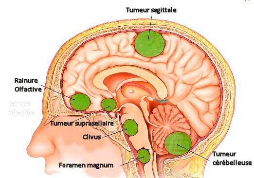
L’étude a révélé que le méningiome sphéno-orbitaire s’est développé chez les femmes ayant reçu un traitement à la progestérone, et la plupart des cas ont été trouvés dans la cinquantaine. Les localisations sphéno-orbitaires comprennent la grande aile sphénoïde avec une extension périorbitaire distincte (Terrier et al., 2018). Le symptôme le plus significatif du méningiome sphéno-orbitaire est une perturbation visuelle dans 95% des cas et pourrait se produire avec une parésie des nerfs oculomoteurs. La chirurgie est nécessaire dans la plupart des cas avec des symptômes, mais comme leur localisation peut rendre difficile une résection complète, une radiothérapie adjuvante est recommandée dans certains cas (Terrier et al., 2018).
This study aimed to analyze the association between the history of the use of hormonal contraceptives and the location of meningiomas in the Indonesian population.
Materials and methods
This study collected retrospective data from medical records of all patients with confirmed meningiomas histologically at the Dr. Sardjito, Yogyakarta, Indonesia, 2019 to early 2021. As the history of hormonal contraception is the main variable of this study, only female patients have been included. In addition, patients without data on the use of hormonal contraceptives have been excluded. Due to the limited data of the medical file concerning the details of the use of hormonal contraception, data on the type of hormonal contraception and the duration of use of contraception was not available. Consequently, we only divided the patients in two groups: those with history of hormonal contraception at any time in their lives and those who do not have such history.
We have also collected data on age, symptoms, results of the pathology (WHO classification and histological type) and MRI or brain scanner before surgery (location and size of meningioma). The measurements of the dimension of the tumor were carried out during the last imaging examination before surgery. Images of Tomodensitometry with contrast and MRI weighted in T1 were used, but in the absence of contrast administration, the measures were still carried out provided that the examiner could identify the limit of the lesion with confidence. After the Recist measurement, the longest diameter of the tumor in the axial plane was recorded. The second diameter perpendicular in the axial plane was also taken to allow the compatibility of the measurement of the tumor according to the WHO. The third longest craniocaudal diameter was taken in the sagittal or coronal plane. The measure included any component of the tumors, that is to say the necrotic zone, the calcification, the cystic component and the intra-dusy part. The size of the tumor was determined by multiplying the two longest diameters on the imaging results.
All data were analyzed using IBM SPSS statistics version 26. A bivariate analysis was carried out to analyze the association between the use of hormonal contraceptives and the location of meningiomas. We used an independent T test or the Mann-Whitney test for digital data and the Test of the Chi square or the exact Fisher test for category data. A multivariate analysis using logistics regression with the retrograde method was then carried out to identify the variables independently associated with the location of meningiomas.
Results
The demographic and clinical characteristics of patients are presented in Table 1. Among the 99 patients included in this study, 72 (72.7 %) had history of hormonal contraception, while 27 (27.3 %) did not.
The average age at the time of the diagnosis of meningioma was 47.92 ± 8.29 years. There was no significant age difference between groups with and without hormonal contraception (p = 0.875).
The subjects consist of 83 (83.8%) OMS grade I and 16 (16.2%) tumors of Grade II and III of WHO. The most frequent histological types were meningothelial (33.3 %) and transitional (24.2 %). There was no statistically significant difference in the WHO classification and the histological type between hormonal contraception groups (p = 0.129 and 0.112, respectively). We did not find a statistical difference between hormonal contraception and the size of the tumor (p = 0.974).
We did not find a difference in the performance scale of Karnofsky (KPS) to the intake between the two groups (p = 0.517). Patients in the hormonal contraception group presented significantly more visual symptoms than those without history of hormonal contraception (66.7 vs 44.4% respectively, p = 0.044).
A bivariate analysis was carried out to analyze the association between various variables and the location of meningiomas (Table 2). A total of 57 (57.6 %) tumors were located in the spheno-organ, and 42 (42.4 %) were distributed in other locations. There was no significant difference between the two groups with regard to age at the time of diagnosis (p = 0.779), the histological type (p = 0.126) and the size of the tumor (p = 0.772).
As expected, patients with spheno-orbital meningioma had more visual symptoms than the group of tumors located in other locations (78.9 % against 35.7 %, p <0.001). In addition, patients with spheno-orbital meningioma also presented less crises (p <0.001) and fewer changes in speech (p = 0.039).
A significant association was found between the rank of meningiomas and their location, with a higher proportion of MENINGIMES of grade I of WHO in spheno-orbital tumors than in the meningiomas of other locations (91.2 vs 73.8%, p = 0.02).
Nous avons trouvé une différence significative dans la localisation des tumeurs entre les groupes de contraception (p=0,038). Les patientes ayant des antécédents de contraception hormonale présentaient une proportion plus élevée de méningiomes dans des localisations sphéno-orbitales que les patientes sans ces antécédents. (63,9 vs. 40,7% respectivement, p=0,038). Une analyse multivariée a ensuite été réalisée pour confirmer ce résultat. Les variables qui pouvaient potentiellement affecter la localisation des méningiomes et dont le p
Table 1. Demographic and clinical characteristics of subjects (n = 99)
| Variable | Total | Hormonal contraception | P-Value | ||||
| No yes | |||||||
| Number of patients, n (%) | 99 (100) | 27 (27.3) 72 (72.7) | 72 (72.7) | ||||
| Average age, years (sd) | 47.92 (8.29) | 48.52 (9.18) | 46.38 (6.86) | 0.875* | |||
| WHO grade, n (%) | |||||||
| Grade i | 83 (83.8) | 20 (74.1) | 63 (87.5) | 0.129** | |||
| Others | 16(16.2) | 7 (25.9) | 9 (12.5) | ||||
| Histological type, n (%) | |||||||
| Meningothelial | 33 (33.3) | 9 (33.3) | 24 (33.3) | 0.112** | |||
| Transitional | 24 (24.2) | 9 (33.3) | 15 (20.8) | ||||
| Fibroblastic | 11 (11.1) | 0 (0) | 11 (15.3) | ||||
| Microcystic | 10 (10.1) | 1 (3.7) | 9 (12.5) | ||||
| Atypical | 13 (13.1) | 5 (18.5) | 8 (11.1) | ||||
| Other | 8 (8.1) | 3 (11.1) | 5 (6.9) | ||||
| Size in CM2, average (SD) | 28.68 (19.06) | 27.66 (16.27) | 29.07 (20.16) | 0.974* | |||
| Symptoms, n (%) | |||||||
| Headache | 82 (82.8) | 24 (88.8) | 58 (80.6) | 0.388** | |||
| View disturbances | 60 (60.6) | 12 (44.4) | 48 (66.7) | 0.044 | |||
| Epilepsy crisis | 21 (21.2) | 8 (29.6) | 13 (18.1) | 0.21 | |||
| Unconsciousness | 8 (8.1) | 2 (7.4) | 6 (8.3) | 1** | |||
| Personality change | 6 (6.1) | 2 (7.4) | 4 (5.6) | 0.663** | |||
| Modification of speech | 15 (15.2) | 6 (22.2) | 9 (12.5) | 0.344** | |||
| Cognitive changes | 6 (6.1) | 2 (7.4) | 4 (5.6) | 0.663** | |||
| Anomaly of the process | 2 (2) | 0 (0) | 2 (2.6) | 1** | |||
| Hemiparisia | 18 (18.2) | 8 (29.6) | 10 (13.9) | 0.084** | |||
| Nausea/vomiting | 6 (6.1) | 2 (7.4) | 4 (5.6) | 0.663** | |||
| KPS in admission, average (SD) | 72.61 (20.37) | 77.14 (17.36) | 69.81 (22.71) | 0.517* | |||
*, Mann-Whitney Test; **, Fisher exact test; ***, independent t-test, other tests use chi square test
Discussion
Des études antérieures ont suggéré que le développement des méningiomes est particulièrement influencé par les hormones sexuelles féminines (Bernat et al., 2015). Notre étude a montré qu’environ 70 % des femmes atteintes de méningiome avaient des antécédents de contraception hormonale. Dans la lignée de la nôtre, une étude cas-témoins antérieure en Suède a trouvé un risque élevé de méningiome avec l’utilisation d’une contraception hormonale (Wigertz et al., 2006). La contraception hormonale la plus utilisée dans cette étude était des injections contenant de la progestérone à haute dose.
These results comply with those of a clinical study conducted in Paris, in France, which revealed that spherorbital meningiomas are preferentially developing in women who follow hormone treatment towards the fifties (APRA et al., 2020).
One of the largest cohort studies in France also revealed that the risk of meningioma was much higher in women treated by cyproterone acetate (ACP), a product of progesterone synthesis, with a cumulative dose of more than 60 g. However, after a year of stopping treatment, the risk of meningioma has decreased significantly (Weill et al., 2021).
Une autre étude de cohorte en français a montré une distribution différente des âges chez les femmes qui prenaient de l’ACP par rapport à la population non-ACP. Les femmes qui prenaient de l’ACP avaient un âge médian au moment de la chirurgie du méningiome inférieur de 14 ans à celui du groupe sans ACP (Champeaux-Depond et al., 2021). Parallèlement, dans notre étude, il y avait une légère différence d’âge moyen entre la population sous contraception hormonale (46,38±6,86 ans) et celle sans contraception hormonale (48,52±9,18 ans). Cependant, la différence n’était pas statistiquement significative.
Previous studies have shown the possible association between the use of hormonal contraception and the location of meningiomas, in particular in the base of the skull (Peyre et al., 2018). APRA (2020) has found that sphero-orbital meningiomas are developing more often in women under hormonal contraception.
L’étude de Champeaux-Depond (2021) a montré que les méningiomes induits par la contraception hormonale étaient préférentiellement situés sur la base antérieure et moyenne du crâne. De même, Peyre (2018) a rapporté une prédominance de la base du crâne antérieure chez les patients atteints de méningiomes ayant pris une contraception hormonale. Dans cette étude, nous avons trouvé un méningiome sphéno-orbitaire dans 57 (57,6%) cas. Statistiquement, nous avons trouvé une association significative entre l’utilisation de contraceptifs hormonaux et la localisation du méningiome dans la région sphéno-orbitaire en analyse bivariée (p=0,038). Une analyse multivariée a confirmé cette association, montrant que les patientes ayant des antécédents de contraception hormonale étaient deux fois et demie plus susceptibles de développer un méningiome dans la région sphéno-orbitaire que les patientes sans antécédents de contraception hormonale (p=0,041). Cependant, le mécanisme sous-jacent n’est pas clairement compris. Cela est probablement lié au niveau plus élevé de récepteurs de la progestérone (PR) des méninges dans cette région. Des études antérieures ont montré que les méningiomes de la base du crâne médiane présentent un nombre significativement plus élevé de cas avec une forte expression de PR (Apra et al.,2020 ; Maiuri et al., 2021). Cependant, il y a un manque de preuves associant la quantité de récepteurs hormonaux à la croissance tumorale sous traitement (Apra et al., 2020). Ce point nécessite une étude plus approfondie.
A greater visual impairment in patients with hormonal contraception of contraception could be linked to the location of the tumor in this spheno-orbital region resulting in compression of the optic nerve. Indeed, our study showed that out of 72 patients with history of hormonal contraception, 48 (66.7%) of them had a visual impairment (p = 0.04). Almost 80 % of patients with spheno-orbital meningioma presented a visual impairment, which is much higher than meningiomas of other locations (35.7 %) (p <0.001). A meta-analysis study had reported that the most present symptoms in sphening-organic meningiomas were proptosis (84%), unilateral visual impairment (46%) and the deficit in the visual field (31%) (Fisher et al., 2021). Consequently, the first -line treatment is surgery due to the compression of the optic nerves (Honeybul et al., 2001). In addition, spheno-orbital meningioma is also known to have a higher recurrence rate than meningiomas in other locations (Terrier et al., 2018).
La principale limite de cette étude est le manque d’informations sur la durée, les doses ou le type de contraception hormonale utilisée. Par conséquent, une étude plus approfondie pour explorer l’association de ces variables avec la localisation des méningiomes dans la population indonésienne est nécessaire. Une autre limite est le petit nombre de méningiomes de grade II et III de l’OMS dans notre étude.
In conclusion, the use of hormonal contraception is associated with meningiomas in the Spheno-Orbital region. The result is a higher number of patients with visual symptoms in the group with history of hormonal contraception.
Table 2. Bivariate analysis of the location of meningiomas
| Variable | Total | Location | P-Value | ||||
| Other spheroorbital | |||||||
| Number of patients, n (%) | 99 (100) | 57 (57.6) | 42 (42.4) | ||||
| Average age, years (sd) | 47.92 (8.3) | 47.89 (8.5) | 47.95 (8.04) | 0.779* | |||
| WHO grade, n (%) | |||||||
| Grade i | 83 (83.8) | 52 (91.2) | 31 (73.8) | 0.02 | |||
| Others | 16 (16.2) | 5 (8.8) | 11 (26.2) | ||||
| Histological type, n (%) | |||||||
| Meningothelial | 33 (33.3) | 22 (38.6) | 11 (26.2) | 0.126** | |||
| Transitional | 24 (24.2) | 14 (24.6) | 10 (23.8) | ||||
| Fibroblastic | 11 (11.1) | 8 (14) | 3 (7.1) | ||||
| Microcystic | 10 (10.1) | 6 (10.5) | 4 (9.5) | ||||
| Atypical | 13 (13.1) | 3 (5.3) | 10 (23.8) | ||||
| Other | 8 (8.1) | 4 (7.1) | 4 (9.5) | ||||
| Size in CM2, average (SD) | 28.68 (19.06) | 28.29 (20.04) | 29.17 (18.03) | 0.772* | |||
| Symptoms, n (%) | |||||||
| Headache | 82 (82.8) | 45 (78.9) | 37 (88.1) | 0.233 | |||
| Visual disturbances | 60 (60.6) | 45 (78.9) | 15 (35.7) | <0.001 | |||
| Epilepsy crisis | 21 (21.2) | 4 (7.1) | 17 (40.5) | <0.001 | |||
| Unconsciousness | 8 (8.1) | 3 (5.3) | 5 (11.9) | 0.278** | |||
| Personality change | 6 (6.1) | 2 (3.5) | 4 (9.5) | 0.397** | |||
| Change of speech | 15 (15.2) | 5 (8.8) | 10 (23.8) | 0.039 | |||
| Cognitive change | 6 (6.1) | 1 (1.8) | 5 (11.9) | 0.08** | |||
| Anomaly of the process | 2 (2) | 1 (1.8) | 1 (2.4) | 1** | |||
| Hemiparisia | 18 (18.2) | 7 (12.3) | 11 (26.2) | 0.076 | |||
| Nausea/vomiting | 6 (6.1) | 2 (3.5) | 4 (9.5) | 0.397** | |||
| KPS in admission, average (SD) | 72.61 (20.37) | 72.45 (21.2) | 72.82 (19.46) | 0.819* | |||
| Use of hormonal contraceptives, n (%) | |||||||
| Yes | 72 (100) | 46 (63.9) | 26 (36.1) | 0.038 | |||
| No | 27 (100) | 11 (40.7) | 16 (59.3) | ||||
*, Mann-Whitney Test; **, Fisher exact test; ***, independent t-test, other tests use chi square
Table 3. Multivariate analysis by logistical regression of factors affecting the location of meningiomas
| Variable | Odds Ratio | 95 % CI | P-Value |
| Use of hormonal contraceptives | 2.573 | 1.040-6.367 | 0.041 |
Author's declaration of contribution
RGM and EKD formulated the idea presented and designed the study. RAH, DNFF, KRD, AR, MAS, ASW, KD, EBS, AA and RGM collected samples and clinical data. NHS analyzed all MRI and CT exams. RGM and EKD have developed the theory, carried out the statistical analysis and wrote the initial version of the manuscript. All the authors discussed the results and contributed to the final manuscript. RGM has prepared the final manuscript. All projects were supervised by EKD.
Thanks
This research was supported by the PDUPT grant from the Indonesian Ministry of Research and Higher Education, number 8/E1/KPT/2021 and 3572/E4/AK.04/2021 in EKD.
Ethical approval
This study obtained the ethical approval of the Institutional Review Board (IRB), Faculty of Medicine, Public Health and Nurses, Universitas Gadjah Mada, Indonesia. The written informed consent was obtained from the patients themselves or a member of their family.
Conflicts of interest
The authors have no conflict of interest to declare.
References
APRA C, Roblot P, Alkhayri A, et al (2020). Female gender and exogenous progesterone exhibition as risk factors for spheno-organ meningiomas. J Neurooncol, 149, 95-101.
Baldi I, Engelhardt J, Bonnet C, et al (2018). Epidemiology of meningiomas. Neurosurgery, 64, 5-14.
Bernat Al, Oyama K, Hamdi S, et al (2015). Growth Stabilization and Regression of Meningiomas after Discontinuization of Cyproterone Acetate: A Case Series of 12 Patients. Acta Neurochir, 157, 1741-6.
Champeaux-Deland C, Weller J, Froelich S, Sartor A (2021). Cyproterone acetate and meningioma: a nationwide-wide population based study. J Neurooncol, 151, 331–8.
Fisher FL, Zamanipoor Najafabadi Ah, Schoones JW, Genders SW, Furth WR (2021). Surgery as a safe and effective Treatment option for spheno-orbital meningioma: A Systematic Review and Meta-Analysis of Surgical Techniques and Outcomes. Acta Ophthalmol, 99, 26-36.
Fisher Fl, Zamanipoor Najafabadi Ah, van der Meer Pb, et al (2021). Long-Term Health-Related Quality of Life and Neurocognitive Functioning After Treatment in Skull Base Meningioma patients. J neurosurg, 1, 1-13.
Goldbrunner R, Stavrinou P, Jenkinson MD, et al (2021). Eano Guideline on the Diagnosis and Management of Meningiomas. Neurooncol, 23, 1821-34.
Harland TA, Freeman JL, Davern M, et al (2018). Progesteroneonly contraception is associated with a shorter progressionfree survival in premenopausal women with who grade i meningioma. J Neurooncol, 136, 327-33.
Honeybul S, Neil-Dwyer G, Lang Da, Evans BT, Ellison DW (2001). Sphenoid Wing Meningioma in plate: A Clinical Review. Acta Neurochir, 143, 749-58.
Korhonen K, Salminen T, Raitanen J, et al (2006). Female predominance in meningiomas can not be explained by different in progesterone, estrogen, or Androgen receptor expression. J Neurooncol, 80, 1-7.
Lee YS, Lee YS (2020). Molecular characteristics of meningiomas. J pathol transl med, 54, 45.
Mauri F, Mariniello G, from Divitiis O, et al (2021). Progesterone Receptor Expression in Meningiomas: Pathological and Prognostic Implications. Front Oncol, 11, 2585.
Ostrom QT, Gittleman H, Truitt G, et al (2018). CBTRUS Statistical Report: Primary Brain and Other Central Nervous System Tumors Diagnosed in the United States in 2011–2015. Neurooncol, 20, IV1-86.
Peyre M, Gaillard S, de Marcellus C, et al (2018). Progestinassocated shift of Meningioma Mutual Landscape. Ann Oncol, 29, 681-6.
Qi Zy, Shao C, Huang Yl, et al (2013). Reproductive and exogenous hormone factors in Relation to risk of meningioma in women: a meta-analysis. PLOS One, 8, E83261.
Terrier LM, Bernard F, Fournier HD, et al (2018). SPHENO-ORBITAL MENINGIOMAS SURGERY: MULTICENTER Management Study for Complex Extensive Tumors. World Neur, 112, E145-56
Van den Munckhof P, Christiaans I, Kenter SB, Baas F, Hulsebos TJM (2012). Germline SMARCB1 PREDISPOSES TO MULTIPLE MENINGIOMAS AND SCHWANNOMAS with preferential rental of Cranial meningiomas at the FALX CEREBRI. Neurogenetics, 13, 1-7.
Weill A, Nguyen P, Labidi M, et al (2021). Use of High dose Cyproterone Acetate and Risk of Intracranial Meningioma in Women: Cohort Study. BMJ, 372.
Wiemels J, Wrensch M, Claus EB (2010). Epidemiology and Etiology of Meningioma. J Neurooncol, 99, 307-14.
Wigertz A, Lönn S, Mathiesen T, et al (2006). Risk of Brain Tumors Associated With Expores To Exogess Female Sex Hormones. Am J Epidemiol, 164, 629-36.